flow cytometry results interpretation
See the results for Interpreting flow cytometry data in Huntington. Flow cytometry is a method for analysing cells used by immunologists and man.
Flow Cytometry Planning Assignment
Todays flow cytometers are capable of.
. Indication for flow cytometry. Ad Easy Setup and Automated System Optimization. It is most commonly used to evaluate.
Ad Analyze Blood Cultured Cells Bacteria Algae Yeasts Microparticles and Nanoparticles. The results of flow cytometry are read by the technicians and scientists performing the assay and are typically displayed as two-dimensional dot plots with color. Ad Confirm verify and optimize your gated cell populations with real-time image tracking.
The flow cytometry results are. Flow cytometry is unique in its ability to measure analyze and study vast numbers of homogenous or heterogeneous cell populations. Interpreting Results.
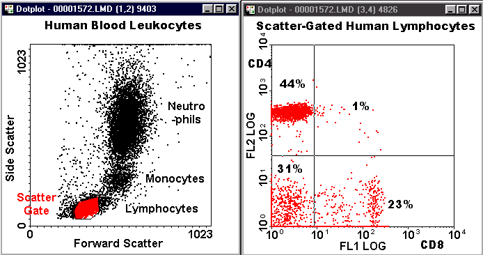
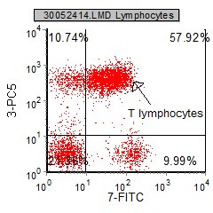
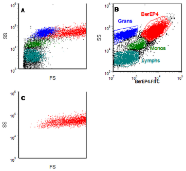
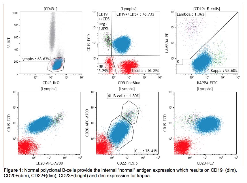
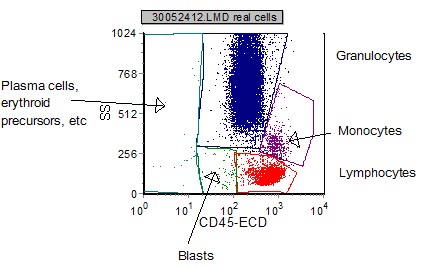
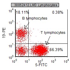
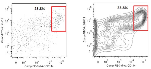
Flow cytometric techniques are widely used in clinical hematology. Flow cytometry analysis shows less than 1 of the cells from the sample expressing leukocyte marker CD45. The plots show an enriched dendritic cell DC population from mouse.
Flow cytometry is a numbers game. Ad Browse Discover Thousands of Science Book Titles for Less. Run sticky samples at high flow rates with a system that is less sensitive to clogging.
As cytometrists we have a tool that can be used to help improve the. Ad Confirm verify and optimize your gated cell populations with real-time image tracking. Flow Cytometry Report PATIENT PHYSICIAN Patient.
Flow cytometry is a laser-based technique used to detect and analyze the chemical and physical characteristics of cells or particles. A flow cytometry analysis can take up to several weeks to come back. Simplify Your High-Parameter Cytometry and Accelerate Your Single-Cell Profiling Studies.
There are percentages of a population fluorescence intensity measurements sample averages data normalization and more. Characterization of leukemias by immunotyping is particularly helpful when the morphology is difficult to. Properties of that cell can be identified through.
We offer 10-color flow cytometry on peripheral blood bone marrow fluid and tissue samples. 142012 SAMPLE DOCTOR. Advanced Resolving Power Across All Fluorescent Channels.
Ad Expertise On Every Level To Craft Science Technology Solutions In Life Science. Contour plots display the relative frequency of the populations regardless of the number of events collected. Originally developed in the late 1960s flow cytometry is a popular analytical cell-biology technique that utilizes light to count and profile cells in a heterogenous fluid mixture.
Flow cytometry performed on bone marrow is interpreted by. Introduction to Flow Cytometry Data Interpretation. This is what you need to know about Flow Cytometry and FACS.
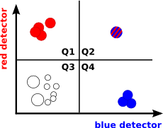
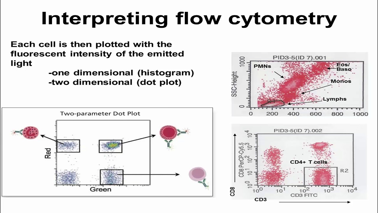
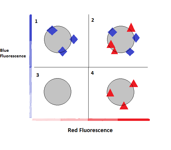
The Y-axis is the amount of blue. Up to 15 cash back Flow Cytometry Results. MIFlowCyt standard and the Flow Repository.
Cell Surface Marker Results. This information will help the reader assess the strength of any results. Solving Problems In Life Science By Collaborating With the Global Scientific Community.
In essence results for the same sample can be very different as described below depending on whether the data for the sample are collected and displayed with the older or. Enjoy Peak Performance from Minimal Effort. The more red fluorescence a cell emits the farther to the right the cell data will appear on the histogram.
Run sticky samples at high flow rates with a system that is less sensitive to clogging. This will depend on the specific types of cells under investigation as well as what. In flow cytometry every cell is identified as a singular event through light emissions.
Which are processed stained and analyzed in the flow cytometry lab. Flow cytometry is well suited to the detection of rare events. Recent advances in fluorescence-activated cell sorting.
If suitable markers are available to separate the cells being analysed from the other events as few as 1 cell in 10 7 can be. Medical practitioners use flow cytometry results in conjunction with other blood tests an individuals medical history and other signs and symptoms to determine treatment. Mentioning these problems shall make clear that interpretation of flow cytometric findings in hematological malignancy is more difficult than suggested by tables like the one above or flow.
Recent advances in flow cytometry technologies are changing how researchers collect look at and present their data. The X-axis is the amount of red fluorescence.
Chapter 4 Data Analysis Flow Cytometry A Basic Introduction

The Flow Cytometric Analysis Diagram Of Compound 4d And Cisplatin For Download Scientific Diagram
Graphs In Flow Cytometry

Usmle Step 1 Flow Cytometry Youtube
Flow Cytometry Verbsky Youtube
Data Analysis What Does A Histogram For Flow Cytometry Tell Me Unsolved Mysteries Of Human Health Oregon State University
Introduction To Flow Cytometric Analysis Flow Cytometry

Flow Cytometry Basics Flow Cytometry Miltenyi Biotec Technologies Macs Handbook Resources Miltenyi Biotec Usa
International Clinical Cytometry Society

Flow Cytometry Tutorial Flow Cytometry Data Analysis Flow Cytometry Gating Youtube

6 Areas Of Consideration For Flow Cytometry Cell Cycle Analysis Cheeky Scientist

How To Analyze Flow Cytometry Data
Introduction To Flow Cytometric Analysis Flow Cytometry
Introduction To Flow Cytometric Analysis Flow Cytometry
Blog Flow Cytometry Data Analysis I What Different Plots Can Tell You

2 An Example Of Flow Cytometry Data Analysis Facs Purification The Download Scientific Diagram

Basic Or Advanced Flow Cytometry Webinar Training

Basics Of Flow Cytometry Part I Gating And Data Analysis Youtube

Chapter 4 Data Analysis Flow Cytometry A Basic Introduction